FREE PARKING - Downtown Victoria - (250) 940-8111

Dental Technology In Victoria, BC
Dental technology has introduced innovative advancements over the last few years, making dental appointments quicker and much more thorough. Some of the laborious tasks of dentistry have been simplified and the process for several of these duties has proven more efficient. Technology has already altered our everyday lives at home and in the workplace, making it only a matter of time until modern developments changed how patients perceived a routine dental appointment. Here are the pieces of technology we have in our office.
-
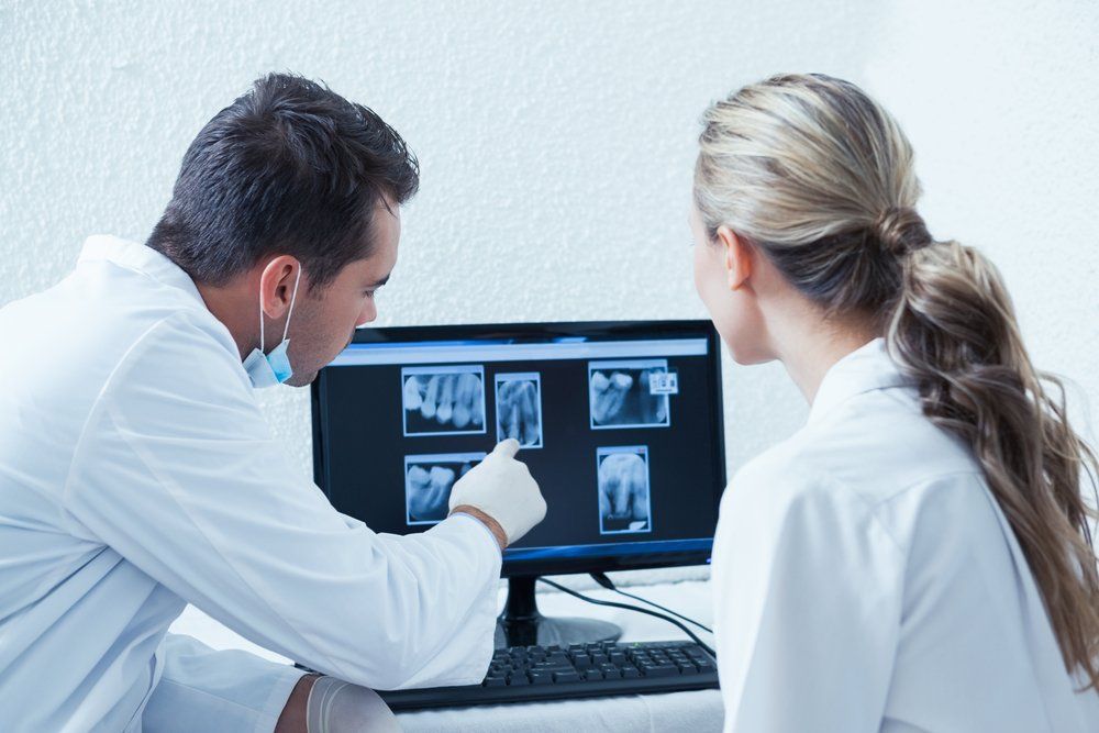
Digital X-Rays
Introduced in 1987, nearly 90 years after traditional x-rays came to fruition, digital radiography combined the power of computer technology with electric sensors and tiny bursts of radiation. Rather than printing the results on film, images form almost as soon as the sensors are placed in our mouths, projecting on a computer screen. Digital x-ray technology does demand additional training for dentists, though the majority of practitioners are adamant that the advantages are worth the commitment. Today, a lot of dental offices only offer patients digital x-rays because, in multiple ways, it is the superior option to traditional radiography.
- Less Expensive | Digital x-rays will generally cost you less than the traditional alternative because the cost of film to develop images for the latter adds up. In contrast, digital x-ray imaging projects right onto our computer.
- Better Storage | Since these digital x-ray images are transferred to a computer system, it allows for easier storage of your oral health records. Your data can be transferred from one dentist to another without any medical data being lost in the exchange.
- Finer Images | Digital x-ray images produce a better resolution than their traditional counterpart. Also, old-fashioned x-rays can only project images in 25 various shades, whereas a digital image can reveal up to 256 shades of grey. Digital radiography also has the advantage of accessing more angles within our mouths, providing a streamlined view of a patient's entire oral structure. With the assistance of computer programs, dentists can even enhance the digital images further, for a focused view.
-
VELscope
VELscope's Vx Enhanced Oral Assessment System is a handheld scope utilized by dentists to help visualize oral tissue abnormalities, including cancer and pre-cancer. This device is used together with, and as a supplement, to the traditional intra and extraoral head and neck examination. The VELscope Vx differs from other adjunctive devices used for oral examination by not requiring any dyes or extended testing procedures. A VELscope Vx exam is completed in our office during a routine hygiene appointment, in roughly two minutes.
Revolutionizing how oral mucosal examinations are executed, the VELscope Vx handheld device emits a bright blue light that is used to inspect our mouths and tongue. As the device is sensitive to abnormal tissue changes, the blue-spectrum light causes the soft tissue (oral mucosa) of our mouths to naturally fluoresce. Healthy tissues glow in distinct patterns that may be visibly disrupted when tissue undergoes an abnormal change, like when it is associated with oral cancer or dysplasia. Potential cancerous or pre-cancerous tissues can be invisible to even a trained eye, which is why the VELscope Vx device is vital in aiding the traditional intra and extraoral head and neck examination. More thorough exams provide our dentist a better chance to identify suspicious areas and quickly investigate for potential oral disease. Such early discovery is imperative because it increases the five-year survival rate for oral cancer patients to about 83 percent.
-
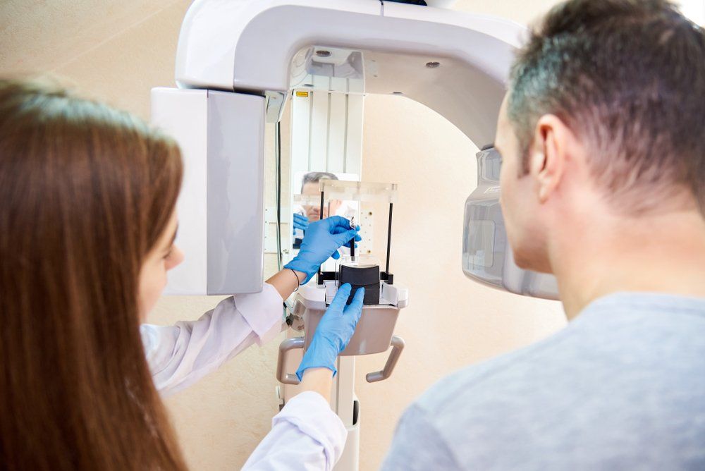
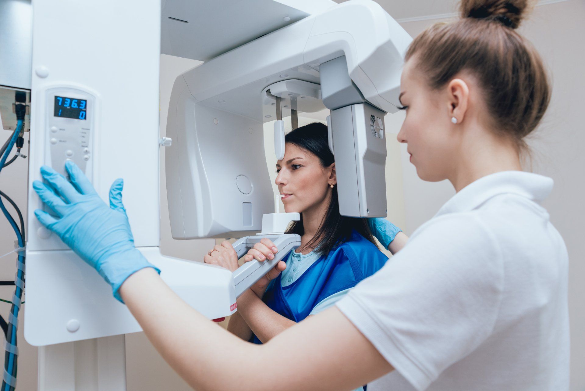
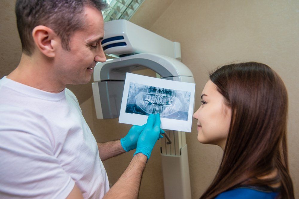
CBCT Machine
Dental cone beam computed tomography (CBCT) is a special type of x-ray machine that is implemented in scenarios where normal dental or facial x-rays are insufficient. This variation of the CT scanner employs a special type of technology to generate 3-D images of dental structures, soft tissues, nerve paths, and bone in the craniofacial area, in one scan. These images allow for more specific treatment planning. The CBCT machine has an x-ray beam, in the shape of a cone, which moves around you to create a large number of high-quality images, or views. It was developed as a means to produce similar images to what a CT provides, though with a significantly smaller and less costly machine that could be situated in an outpatient office. Providing detailed images of the bone, the CBCT machine evaluates diseases of the jaw, dentition, bony structures of the face, sinuses, and nasal cavity. One shortcoming is that it does not provide the comprehensive diagnostic information available with conventional CT, especially in the analyzing of soft tissue structures such as muscles, glands, nerves, and lymph nodes. The CBCT machine can also be used for reconstructive surgery, cephalometric analysis, locating the origin of pain or pathology, surgical planning for impacted teeth, diagnosing temporomandibular joint disorder (TMJ), and for the accurate placement of dental implants.
-
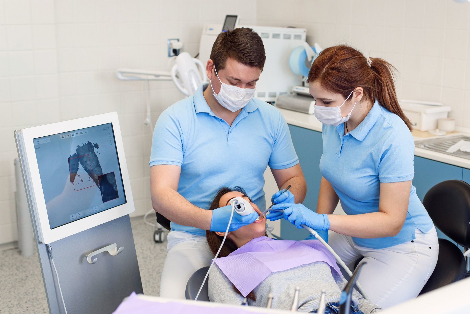
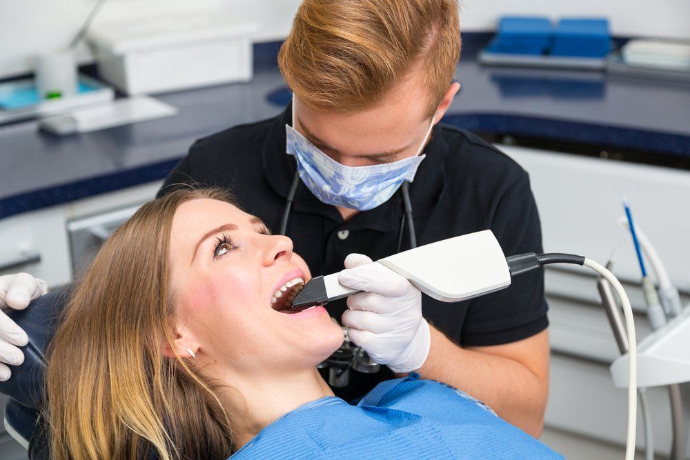
Intraoral Camera
Intraoral cameras are digital imaging tools that dentists utilize to create high-resolution images of your teeth and mouth. These small devices can comfortably fit in your mouth and can be maneuvered over and around your teeth. Once the images are captured, software works to piece them together to develop a digital 3-D model of your mouth. We implement these cameras for various purposes, including a single tooth that needs a restorative procedure or to capture images of soft tissues in our mouths, including our gums and soft palate. A large number of dental issues and conditions that affect our teeth and soft tissues are preventable, which is why intraoral cameras are particular helpful. Dentists can pinpoint and show patients precisely where their oral care needs to be concentrated. Intraoral cameras also limit your time in the office because the images are produced in real-time, and the outcomes are available almost immediately.
-
DEXIS CariVu
DEXIS CariVu is the newest form in digital imaging. It provides patients with a safe and complete oral impression without the use of radiation. The use of DEXIS CariVu is also non-invasive and quite simple. The technology allows our patients to be in-and-out of the office in a few short minutes. This piece of equipment is environmentally friendly too, meaning no films, chemicals, or disposals of molds. We receive instant digital images of your full mouth which helps speed along your treatment plans. Experience the joys of living in the digital age.
-
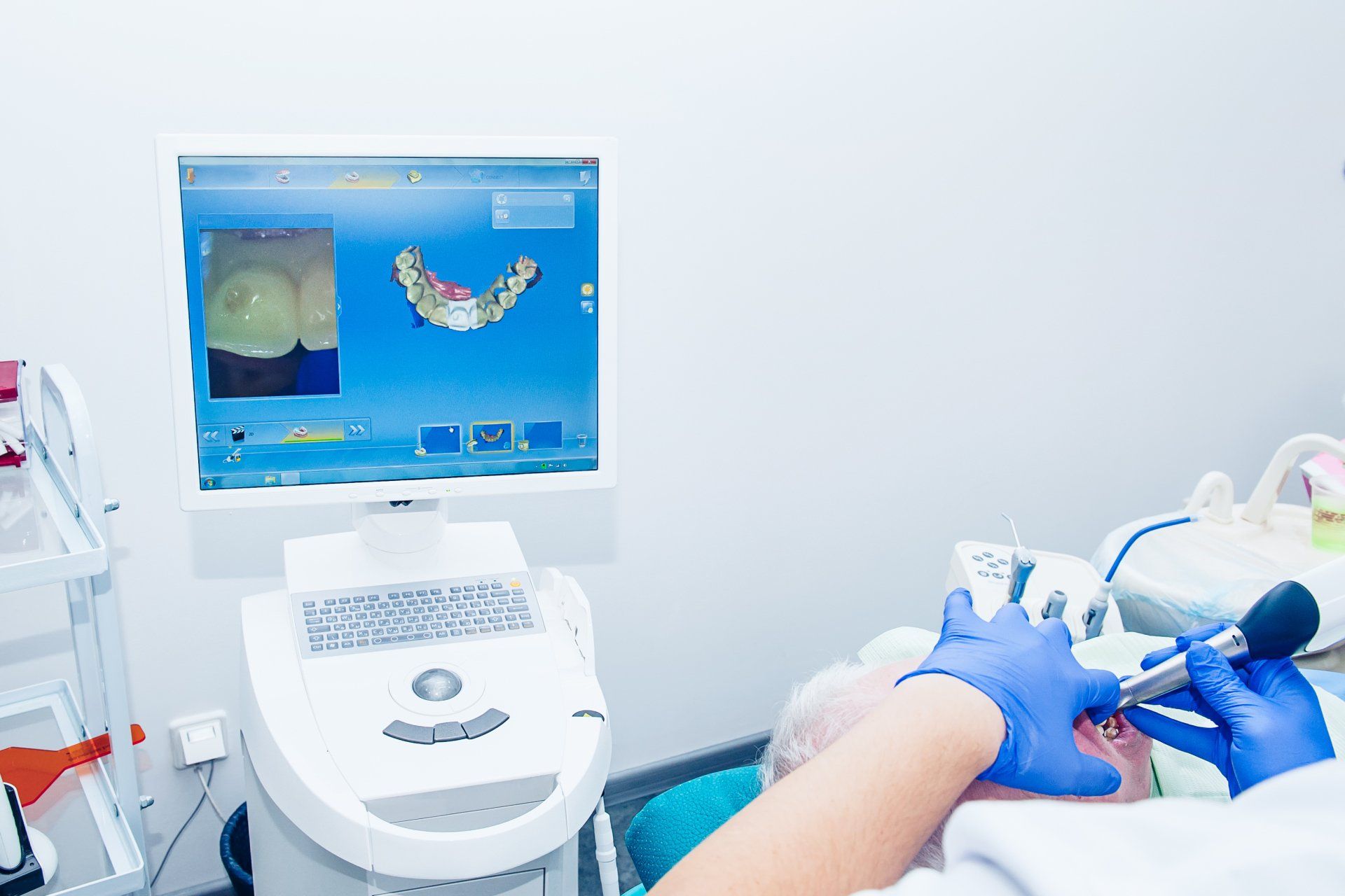
Digital Scanner (iTero)
In 2007, the dental industry made another advancement with a new piece of technology called the iTero digital scanner. This piece of technology takes highly accurate 3-D scans of your mouth. The iTero digital scanner is commonly used to help dental professionals create accurate models for dental restorations which include implants, crowns, and veneers. It is also commonly used for orthodontics to identify issues that may occur during an orthodontic treatment, such as for braces or Invisalign.
-
ARES Sleeping Test
Assessing sleep disorders requires our dentist to accumulate as much data as possible before recommending a treatment plan. This information includes factors that cannot be determined when a patient is awake, such as sleeping position, snoring volume, and other indicators of various sleep disorders. The Apnea Risk Evaluation System (ARES) is an intuitive device utilized to measure several symptoms that patients exhibit in their sleep. First, an ARES questionnaire is provided to patients and the data from this survey looks at snoring history, ethnic background, sleep quality, and more to conclude if a home sleep test is a suitable method of testing. If so, we explain the instructions and demonstrate how you put the unit on before going to sleep. A sensor touches the forehead, and two hose nodules connect to the nostrils, detecting your breathing throughout the night. The ARES test monitors your blood oxygen saturation, nasal airflow, pulse rate, heart rate, and snoring level. At any point, if the sensors or nodules dislodge, the device will attempt to wake you up with a repetitive, loud auditory signal. After you have completed the sleep test, our dentist will analyze the data and determine if further tests or a treatment plan are required.
-
Dental Lasers
Diode Laser | The diode laser is useful for procedures that involve soft tissues, and are great for sterilizing endodontic canals, treating periodontal disease, and teeth whitening. It is a tool that offers a wide array of clinical treatment possibilities and is capable of great precision thanks to its portability and touchscreen controls. It has been shown to be helpful in treating challenging periodontal conditions while providing rapid healing and reduced swelling.
BIOLASE Epic X Laser | BIOLASE Epic X is a state-of-the-art and well-conceived example of a dental diode laser. A variety of pre-set procedures are installed into the device, with suggested settings that can be accessed swiftly and easily by our dentist. The handpiece is fitted with several single-use tips to access any area of our mouths, and there is a handpiece to be used for bio stimulation in pain relief and a whitening contour handpiece for patients who wish to receive laser-assisted in-office whitening. Most commonly, the tips of preference are the ones used intraorally since they are disposable and adjustable to any angle.
-
OccluSense
The OccluSense system was developed to combine the traditional and digital registration of the pressure distribution of occlusal surfaces. Applied in the same manner as conventional occlusion test foils, the OccluSense works in unison with the OccluSense sensors, digitally recording a patients masticatory pressure distribution in 256 pressure levels. Afterwards, the information is transferred to the OccluSense iPad app for further examination and the recordings are stored in our patient management system, ready to be reviewed or exported instantly. OccluSense's Electronic Pressure Sensor is 60 microns thin, flexible, and color-coated, as the material permits the recording of both static and dynamic occlusion. Also, the red color-coated sensor marks the occlusal contacts on a patient's teeth. Where this system separates itself from the traditional occlusion test materials (papers, foils) is that it reveals the local premature contacts on the occlusal surfaces of our teeth, as well as a graphical representation of occlusal pressure conditions, right on the iPad. It is now possible to visualize the proportion of the masticatory forces of the entire dental arch and provide support for patient-oriented therapy planning. Recordings from before and after the dental treatment can be compared and saved for documentation and future reference.
-
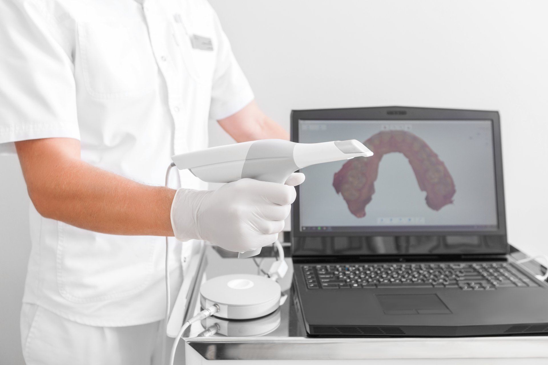
3Shape Digital Wand (Trios)
Digital dentistry has taken its next step with the 3Shape Intraoral Trios Scanner, which is used to create a digital impression of a tooth's anatomy and the tri-dimensional position of dental implants. Information is electronically stored, and this form of digital dentistry affords patients the convenience of not having to sit through traditional impressions, with their unsavory tasting materials, bulky trays, and possible gagging effects. Around five percent of dentists worldwide currently use intraoral scanning but having the capabilities to make a replica of an entire mouth in less than two minutes, that statistic should only grow over time. Traditional methods of taking dental impressions, referred to as "analog", simply cannot keep up with the digital alternative. Digital imaging from the 3Shape Intraoral Trios Scanner is near flawless, an amazingly accurate tool for creating dental crowns, implants, and orthodontic aligner trays. As if that was not enough of an endorsement, the scanner also functions as a camera and operates at remarkable speeds.
-
Air-Filtration (Surgery Clean Air)
We understand that staying inside can cause air to become dense. As more people filter through an area, it is common that the air quality may lack in cleanliness. Due to the constant change in temperature, a building may hold onto bacteria, mold, or illnesses. However, we have an air-filtration system, commonly known as Surgically Clean Air. It is an air cleaning process that has a six-step filtration which helps remove any harmful bacteria in the surrounding area. Our patient's health is of the utmost importance to us, which is why we have implemented this system in our office.
We are the local Victoria Dentist near you!
Rediscover your confidence and your smile
Request Your Next Dental Appointment
We look forward to seeing you soon!
Please note, we will try our best to accommodate your schedule
Thank you so much for contacting our dental practice. While we strive to respond to all inquiries right away, we may be away from the desk helping a patient or out of the office. We will do our best to reach out to you shortly.
Please note, if this is a dental emergency, it would be best to call our practice as this is the fastest way to reach us (250) 940-8111.
Please try again later
Victoria Dentist
We understand that trying to find a nearby dentist you can trust is difficult, that is why we make it easy for you to work with us.
(250) 940-8111
1025 Johnson Street - Unit 401 - FREE Parking
reception@elementsdental.ca
Helpful Links
Dental Practice Hours
- Monday
- -
- Tuesday
- -
- Wednesday
- -
- Thursday
- -
- Friday
- -
- Saturday
- Appointment Only
- Sunday
- Closed
All Rights Reserved | Elements Dental
All Rights Reserved | Elements Dental
Dentist Website Diagnosed, Treated, and Cured by Dr. Marketing Inc